 Csaba Cserháti works at the Department of Solid State Physics, University of Debrecen (Hungary) as an associate professor.
He graduated at the University of Lajos Kossuth University (Debrecen, Hungary) in 1989 he received PhD in Physics there. in 1995. He habilitated in Physics at the University of Debrecen in 2009 and obtained the Doctor of Science title at the Hungarian Academy of Sciences in 2020.
His research field involves investigation of surface and interfacial segregation by computer simulation. Experimental study of solid state reaction on different scale, analysis of nanometer thin film systems by diffraction methods. Interdiffusion studies on a micro and nanoscale; investigation of the Kirkendall effect and its accompanying phenomena. Diffusion in multicomponent alloys as well as atom movements in 2D and 3D metal oxide and metal structures.
Selected topics:
Material transport in different geometry on the nanoscale
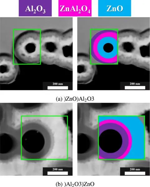
Applying scanning electron microscopy in transmission mode and grazing incidence X-ray diffraction, we investigated the spinel-forming solid-state reaction between crystalline ZnO and amorphous Al2O3 layers in 2D and 3D prepared by atomic layer deposition. In the planar case, we observed two-stage phase growth of the crystalline ZnAl2O4 product layer. During the first stage, flat, pancake-like islands are formed in the nucleation process and these nuclei grow laterally without much increment in thickness. After these islands grow together to form a continuous layer, planar growth is happening in the second stage.
In the 3D cylindrical geometry, two different layer sequences have been studied. To follow the growth kinetics of the spinel, thin lamellae were prepared from the cross sections of the tubes and studied in a transmission mode scanning electron microscope (TSEM). The phase grows parabolically with time, independently of the layer sequence. The growth rate was higher in the ZnO/Al2O3 layer sequence, then for the reverse case. We explained it by the competing vacancy fluxes induced by mechanical stress developing during the solid-state reaction and by the difference in the diffusion fluxes of the two constituents. This is the first case that the influence of closed geometry and layer sequence on spinel growth between two oxide layers has been experimentally investigated.

Vecsei, Gergő; Jáger, Gabriella; Juhász, Laura; Tomán, János J.; Odhiambo, Vincent Otieno; Szilágyi, Imre Miklós; Erdélyi, Zoltán; Cserháti, Csaba
Effect of stacking order in cylindrical geometry on the growth of ZnAl2O4 spinel phase
MATERIALIA 30 Paper: 101819 , 5 p. (2023)
Jáger, Gabriella; Tomán, János J.; Juhász, Laura; Vecsei, Gergő; Erdélyi, Zoltán; Cserháti, Csaba
Nucleation and growth kinetics of ZnAl2O4 spinel in crystalline ZnO – amorphous Al2O3 bilayers prepared by atomic layer deposition
SCRIPTA MATERIALIA 219 Paper: 114857 (2022)
Porous gold nanoparticles
 Cross section view of TPGN, b) Au elemental distribution, c) Ti elemental distribution, d) Au and Ti distribution.") Porous nanoparticles (PGN) are very popular because of their high surface/volume ratio, moreover they have stronger plasmonic properties than their solid counterparts. Due to these properties these are potential candidates in optical, or even in ophthalmological applications. We prepared porous gold nanoparticles on SiO2/Si as well as on sapphire substrates with solid state dewetting – dealloying methods. We studied the morphological and optical properties of PNGs coated with thin (∼7 nm) TiO2 layer using plasma-enhanced atomic layer deposition (PE- ALD) method. Heat-treatments can simply used to tune the optical properties of titania coated porous gold hybrid nanoparticles in a wide range of wavelengths. Changing the composition of the coating layer, that is the mixture ratio, the optical properties can be shifted practically continuously in a wide range determined by the refractive index of the used oxides. As a demonstration, PGNs were coated with mixed alumina-titania oxide layers (5-7 nm) using PE-ALD method. The oxide layer, as an optical tuning tool, also stabilizes the structure of the PGNs when are exposed to elevated temperature. The figure shows a titania coated porous gold nanoparticle annealed at 350 oC for 1 hour. Cross section was prepared by FIB and imaged by TEM/EDS. a) Cross section view of TPGN, b) Au elemental distribution, c) Ti elemental distribution, d) Au and Ti distribution.
Porous nanoparticles (PGN) are very popular because of their high surface/volume ratio, moreover they have stronger plasmonic properties than their solid counterparts. Due to these properties these are potential candidates in optical, or even in ophthalmological applications. We prepared porous gold nanoparticles on SiO2/Si as well as on sapphire substrates with solid state dewetting – dealloying methods. We studied the morphological and optical properties of PNGs coated with thin (∼7 nm) TiO2 layer using plasma-enhanced atomic layer deposition (PE- ALD) method. Heat-treatments can simply used to tune the optical properties of titania coated porous gold hybrid nanoparticles in a wide range of wavelengths. Changing the composition of the coating layer, that is the mixture ratio, the optical properties can be shifted practically continuously in a wide range determined by the refractive index of the used oxides. As a demonstration, PGNs were coated with mixed alumina-titania oxide layers (5-7 nm) using PE-ALD method. The oxide layer, as an optical tuning tool, also stabilizes the structure of the PGNs when are exposed to elevated temperature. The figure shows a titania coated porous gold nanoparticle annealed at 350 oC for 1 hour. Cross section was prepared by FIB and imaged by TEM/EDS. a) Cross section view of TPGN, b) Au elemental distribution, c) Ti elemental distribution, d) Au and Ti distribution.
L Juhász, B Parditka, P Petrik, C Cserháti, Z Erdélyi: Continuous tuning of the plasmon resonance frequency of porous gold nanoparticles by mixed oxide layers Journal of Porous Materials 27 (6), 1583-1588 (2020)
Laura Juhász, Bence Parditka, Shenouda Shanda Shenouda, Misumi Kadoi, Kei-ichi Fukunaga, Zoltán Erdélyi, Csaba Cserháti: Morphological and in situ local refractive index change induced tuning of the optical properties of titania coated porous gold nanoparticles Journal of Applied Physics 128 (5), 054303 (2020)
Laura Juhász, Krisztián Moldován, Petra Herman, Zoltán Erdélyi, István Fábián, József Kalmár, Csaba Cserháti:
Synthesis and Stabilization of Support-Free Mesoporous Gold Nanoparticles Nanomaterials 10 (6), 1107 (2020)
Solid state reaction on the nanoscale
 a room temperature deposited and (b) annealed at 493 K for 30 min.") Evolution of the reaction zone on the nanoscale has been studied in bi- and multilayered Co/a-Si as well as in trilayered Co/a-CoSi/a-Si and Co/CoSi/a-Si thin film diffusion couples. The kinetics of the phase boundary movement during solid state reaction has been followed with special interest of the initial stage of the diffusion, i.e. effects happening on the nanoscale (short time, short distance). The interfacial reactions have been investigated in situ by synchrotron radiation. The formed phases were also characterized by transmission electron microscopy and resistance measurements. The effect of phase nucleation and shift of phase boundaries have been separated in order to determine the “pure” growth kinetics of the crystalline CoSi and Co2Si product phases at the very early stages. Deviations have been found from the traditional diffusion controlled parabolic phase growth. Computer simulations based on a kinetic mean field model illustrated that the diffusion asymmetry (large difference in diffusion coefficients of the materials in contact) may offer a plausible explanations for this. The figure shows TEM images of multilayered sample (a) a room temperature deposited and (b) annealed at 493 K for 30 min.
Evolution of the reaction zone on the nanoscale has been studied in bi- and multilayered Co/a-Si as well as in trilayered Co/a-CoSi/a-Si and Co/CoSi/a-Si thin film diffusion couples. The kinetics of the phase boundary movement during solid state reaction has been followed with special interest of the initial stage of the diffusion, i.e. effects happening on the nanoscale (short time, short distance). The interfacial reactions have been investigated in situ by synchrotron radiation. The formed phases were also characterized by transmission electron microscopy and resistance measurements. The effect of phase nucleation and shift of phase boundaries have been separated in order to determine the “pure” growth kinetics of the crystalline CoSi and Co2Si product phases at the very early stages. Deviations have been found from the traditional diffusion controlled parabolic phase growth. Computer simulations based on a kinetic mean field model illustrated that the diffusion asymmetry (large difference in diffusion coefficients of the materials in contact) may offer a plausible explanations for this. The figure shows TEM images of multilayered sample (a) a room temperature deposited and (b) annealed at 493 K for 30 min.
Z Erdélyi, C Cserháti, A Csik, L Daróczi, GA Langer, Z Balogh, M Varga, DL Beke, I Zizak, A Erko: Nanoresolution interface studies in thin films by synchrotron x‐ray diffraction and by using x‐ray waveguide structure X‐Ray Spectrometry: An International Journal 38 (4), 338-342 (2009)
C Cserháti, Z Balogh, A Csik, GA Langer, Z Erdélyi, Gy Glodán, GL Katona, DL Beke, I Zizak, N Darowski, E Dudzik, R Feyerherm: Linear growth kinetics of nanometric silicides in Co/amorphous-Si and Co/CoSi/amorphous-Si thin films Journal of Applied Physics 104 (2), 024311 (2008)
Hollow-core nanostructures
 and at 723K (diamonds).") Nanoshell formation has been studied experimentally in Ag/Au and Ag/Pd systems in a hemispherical geometry at different temperatures. The void formation in these systems is the result of pure Kirkendall-porosity formation, because it is caused mainly by the inequality of the intrinsic atomic fluxes and other effects (e.g. stresses), inevitably present during nanoshell formations in solid state reactions (oxides, sulphides), can be less important or can be neglected. The kinetics of the process was followed by Transmission Electron Microscopy. Both the growth and shrinkage regimes of the process were observed at the same temperature and even the temperature dependence of the characteristic time (tcr) describing the crossover of the two different regimes was observed. We succeeded to show that tcr shifts to smaller values with increasing temperature. This confirms the theoretical results: the growth and the shrinkage regimes are controlled by the faster as well as the slower diffusion coefficients (DA as well as DB), respectively. It is also illustrated that, confirming recent theoretical predictions, the pore radius linearly depends on the initial particle radius and the slope of this straight line increases with the average composition of the faster component. The plot displayes the relative area of the pores as a function of the annealing time at 743K (squares) and at 723K (diamonds).
Nanoshell formation has been studied experimentally in Ag/Au and Ag/Pd systems in a hemispherical geometry at different temperatures. The void formation in these systems is the result of pure Kirkendall-porosity formation, because it is caused mainly by the inequality of the intrinsic atomic fluxes and other effects (e.g. stresses), inevitably present during nanoshell formations in solid state reactions (oxides, sulphides), can be less important or can be neglected. The kinetics of the process was followed by Transmission Electron Microscopy. Both the growth and shrinkage regimes of the process were observed at the same temperature and even the temperature dependence of the characteristic time (tcr) describing the crossover of the two different regimes was observed. We succeeded to show that tcr shifts to smaller values with increasing temperature. This confirms the theoretical results: the growth and the shrinkage regimes are controlled by the faster as well as the slower diffusion coefficients (DA as well as DB), respectively. It is also illustrated that, confirming recent theoretical predictions, the pore radius linearly depends on the initial particle radius and the slope of this straight line increases with the average composition of the faster component. The plot displayes the relative area of the pores as a function of the annealing time at 743K (squares) and at 723K (diamonds).
G Glodán, C Cserháti, DL Beke: Temperature-dependent formation and shrinkage of hollow shells in hemispherical Ag/Pd nanoparticles; Philosophical Magazine 92 (31), 3806-3812 (2012)
G Glodán, C Cserháti, I Beszeda, DL Beke: Production of hollow hemisphere shells by pure Kirkendall porosity formation in Au/Ag system; Applied Physics Letters 97 (11), 113109 (2010)
Al tracer diffusion coefficient in Ni3Al
Interdiffusion experiments were performed between different Ni–Al two-phase alloys. A method is given to estimate the tracer diffusion coefficient of one of the constituents in a binary alloy knowing the tracer diffusion coefficient of the other species. The procedure is based on the so-called Darken-Manning analysis. The tracer diffusion coefficient of Al was determined in this way in Ni3Al:

Our results are compared with others available in the literature. The values for the self diffusivity of Al calculated from inter- diffusion experiments give consistent result. All these measurements support the theory that diffusion of the minor element occurs through a vacancy mechanism in Ni3Al.
 are located in the formed Ni3Al phase.")

Back-scattered electron image of the diffusion zone after annealing at 1000 oC for 196 h. The Kirkendall markers (ThO2 particles) are located in the formed Ni3Al phase.
The Al tracer diffusion coefficients are represented in an Arrhenius-plot. For comparison the Ni tracer diffusion data are represented as well. The error of the tracer diffusion coefficients were calculated with the Gauss’s equation for error propagation using the uncertainties of the experi- mental measurements.
C Cserháti, A Paul, AA Kodentsov, MJH Van Dal, FJJ Van Loo: Intrinsic diffusion in Ni3Al system Intermetallics 11 (4), 291-297 (2003)
Periodic layer formation
 Formation of diffusion zone morphologies periodic in time and space is considered as a manifestation of the Kirkendall effect accompanying reactive diffusion in the solid state. The patterning during multiphase diffusion is attributed to diverging vacancy fluxes within the interaction zone. This generates multiple Kirkendall planes, which by attracting in situ-formed inclusions of the secondary-formed phase can result in a highly patterned structure. Back-scattered electron image of the reaction zone of the Ni50Co20Fe30-Mg diffusion pair at 733 K, 72 h, after heat treatment in Aris show non the left. Morphology of the diffusion zone close to the boundary of the base alloy. Jv, JMg and JNi denotes the diffusion flux of the vacancies, the Mg atom sas well as the Ni atoms. (M is the Matano plane, K1 and K2 are the in-situ formed Kirkendall planes.
AA Kodentsov, MJH van Dal, C Cserháti, A Gusak, FJJ van Loo: Patterning in reactive diffusion; Defect and Diffusion Forum 194, 1491-1502 (2001)
Kirkendall-effect
In a diffusion-controlled interaction, the Kirkendall plane, as marked by inert particles placed at the original contact surface of a reaction couple, need not be unique. Multiple planes can develop, and sometimes the Kirkendall plane does not exist at all. A phenomenological approach is introduced to rationalize the Kirkendall-effect-mediated migration of macroscopic inclusions inside a diffusion zone.
 of the diffusion zone after interaction.")

Back-scattered electron image (BEI) of the diffusion zone after interaction on the left. On the right panel the Kirkendall velocity curve corresponding to the β’-AuZn product layer in the annealed (500°C; 17.25h; Ar+5vol.% H2) Au/Au36Zn64 (β-’AuZn2’) couple constructed on the basis of the positions of the Kirkendall planes and the point where v≈0 (sudden change in contrast) relative to the phase boundaries. Three intersections between the velocity curve and the line v= x/2t (K1, K2 and K3) are found, of which only two correspond to stable Kirkendall planes (at K1 and K3). (The position x =0 represents the Matano plane.)
MJH Van Dal, AM Gusak, C Cserháti, AA Kodentsov, FJJ van Loo: Microstructural Stability of the Kirkendall Plane in Solid-State Diffusion; Physical Review Letters 86 (15) (2003)